Renowned Japanese dentist Dr. Hidetaka Ishizaki recently provided detailed feedback on two clinical cases where he used Rogin’s SF-shape rotary file for root canal treatments. His insights emphasize the effectiveness and precision of the SF-shape rotary file in endodontic procedures. Here, we delve into the specifics of these cases to highlight the capabilities of this advanced dental tool.
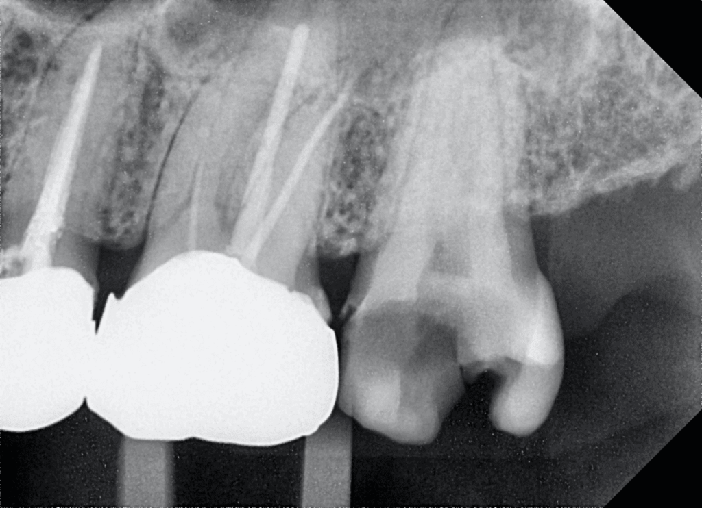
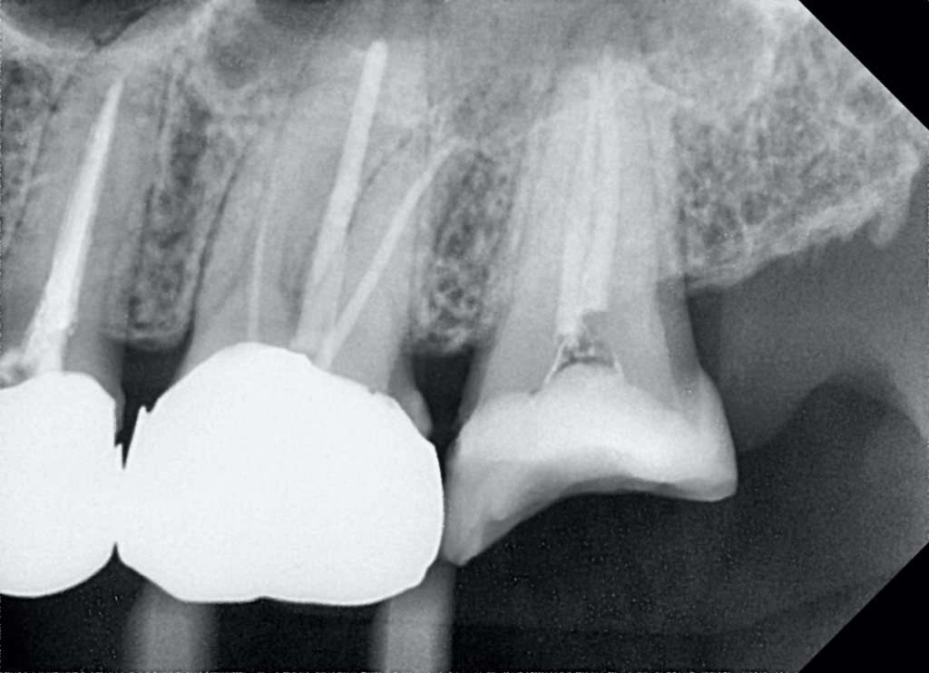
Case 1: Maxillary Left Second Molar
Patient Profile
– Age/Gender: 40-year-old female
– Symptoms: Biting pain on maxillary left second molar, tender to percussion, no spontaneous pain.
– Diagnosis: Large caries with exposed pulp
Procedure
Preoperative Steps: Removal of caries and temporary buildup with flowable resin.
Canal Preparation:
– Instruments Used: TORNA SF-shape rotary files
– Steps:
– Enlargement of root canal orifices with the orifice opener of the SF-shape rotary files.
– Working length measurement followed by canal preparation up to 25/.04 with the SF-shape rotary files.
– Irrigation: 2.5% NaClO solution.
Filling and Restoration:
– Filling: Size 25/.04 Gutta percha and CSBS.
– Restoration: Tooth was built up with a fiber core and restored with a metal crown.
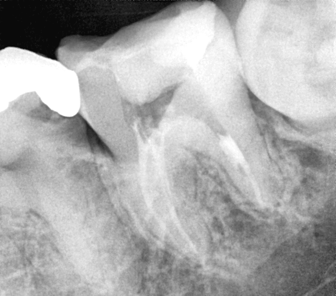
Case 2: Mandibular Left Second Molar
Patient Profile
– Age/Gender: 27-year-old male
– Symptoms: Spontaneous pain at left lower molar region, unable to sleep due to pain, responsive to percussion, positive to electrical pulp test.
– Diagnosis: Deep caries at the distal aspect, vital pulp not exposed initially.
Procedure
Initial Treatment: Indirect pulp capping with MTA putty.
– Outcome: No improvement in symptoms, leading to a decision for pulpectomy.
Canal Preparation:
– Instruments Used: TORNA SF-shape rotary files
– Steps:
– Access opening revealed necrotic coronal pulp.
– Preparation of thin and curved mesio-buccal and mesio-lingual root canals, and oval distal root canal.
– Used SF-shape rotary files 16, 20/.04, and 25/.04 for distal root canal preparation.
– Measurement and Patency: Apex locator used for working length measurement. Preparation confirmed apical patency and maintenance of original root canal morphology.
– Irrigation: 2.5% NaClO solution with each file change.
Filling:
– Mesio-buccal and Mesio-lingual Canals: Filled with 20/.04 Gutta percha.
– Distal Canal: Filled with 25/.04 Gutta percha and CSBC as a single cone.
Conclusion
Dr. Hidetaka Ishizaki’s feedback on using the SF-shape rotary file underscores its efficiency in root canal preparation and its ability to maintain the original canal morphology. These cases illustrate the reliability and precision of the SF-shape rotary file in managing complex endodontic treatments. The advanced design of the SF-shape rotary file not only facilitates smooth and efficient canal preparation but also ensures patient comfort and successful outcomes.
By integrating the SF-shape rotary file into clinical practice, dental professionals can enhance the quality of endodontic treatments and achieve superior results, as demonstrated in these cases by Dr. Hidetaka Ishizaki.
CHINA LEADING MANUFACTURER OF ENDODONTIC INSTRUMENTS
Please leave your contact information so that we can contact you as soon as possible
Ask For A Quick Quote
We will contact you within 1 working day, please pay attention to the email with the suffix “@rogindental.com”.
Kind tipsRotary files: Over 500 packs
Hand use files: Over 1000 packs
Equipment:
Large machines: 2+ units
Motors: 3+ units
If your order meets these requirements, please mark your status as "Dealer" when submitting your inquiry or order. Thank you!