Abstract
Separated endodontic instruments remain one of the most challenging complications in root canal retreatment. Successful management requires excellent visualization, conservative techniques, and advanced instrumentation to retrieve the fragment while preserving dentin and achieving thorough disinfection and obturation.
This case report presents the non-surgical retreatment of tooth #14 (maxillary first premolar) in a patient with symptomatic apical periodontitis due to a failed previous root canal treatment. A 2 mm separated instrument lodged in the apical third of the buccal canal was successfully retrieved using a dental operating microscope and an ultrasonic E6 tip. Retreatment was completed in a single visit using Rogin rotary retreatment files for material removal, Rogin Super Flexi files for shaping, an activated irrigation protocol, and 3D obturation with bioceramic sealer. The treatment resulted in complete resolution of symptoms and a favorable clinical outcome.
Introduction
Instrument separation during endodontic treatment or retreatment can prevent adequate cleaning, shaping, and obturation of the apical region, often leading to persistent infection and symptomatic apical periodontitis. Retrieval of separated instruments, especially when located in the apical third, demands high magnification, precise ultrasonic application, and minimal dentin removal to avoid perforation or weakening of the root structure.
Modern tools—such as the dental operating microscope, specialized ultrasonic tips, flexible NiTi rotary files, activated irrigation, and bioceramic sealers—have significantly improved the predictability of such complex retreatments. This report describes a single-visit retreatment of tooth #14 involving retrieval of a 2 mm separated file fragment, followed by complete biomechanical preparation and 3D obturation.
Case Presentation
Patient History and Clinical Examination
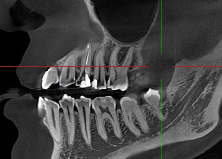
A patient presented with pain on biting in the maxillary right posterior region. Tooth #14 (maxillary first premolar) had a history of previous root canal treatment. Clinical examination revealed tenderness to percussion, particularly during mastication. No swelling or sinus tract was present.
Radiographic evaluation confirmed inadequate previous obturation and a separated instrument fragment visible in the apical third of the buccal canal.
Diagnosis and Treatment Plan Diagnosis:
Symptomatic apical periodontitis secondary to failed root canal treatment with a separated instrument.
Treatment Plan:
- Non-surgical endodontic retreatment
- Removal of previous obturation material
- Retrieval of the 2 mm separated instrument using microscope and ultrasonic instrumentation
- Biomechanical preparation of the root canal system
- 3D disinfection using activated irrigation
- Obturation with bioceramic sealer and 3D technique
- Completion in a single visit if canals remained dry and symptom-free
Clinical Procedure
Access and Retrieval of Separated Instrument
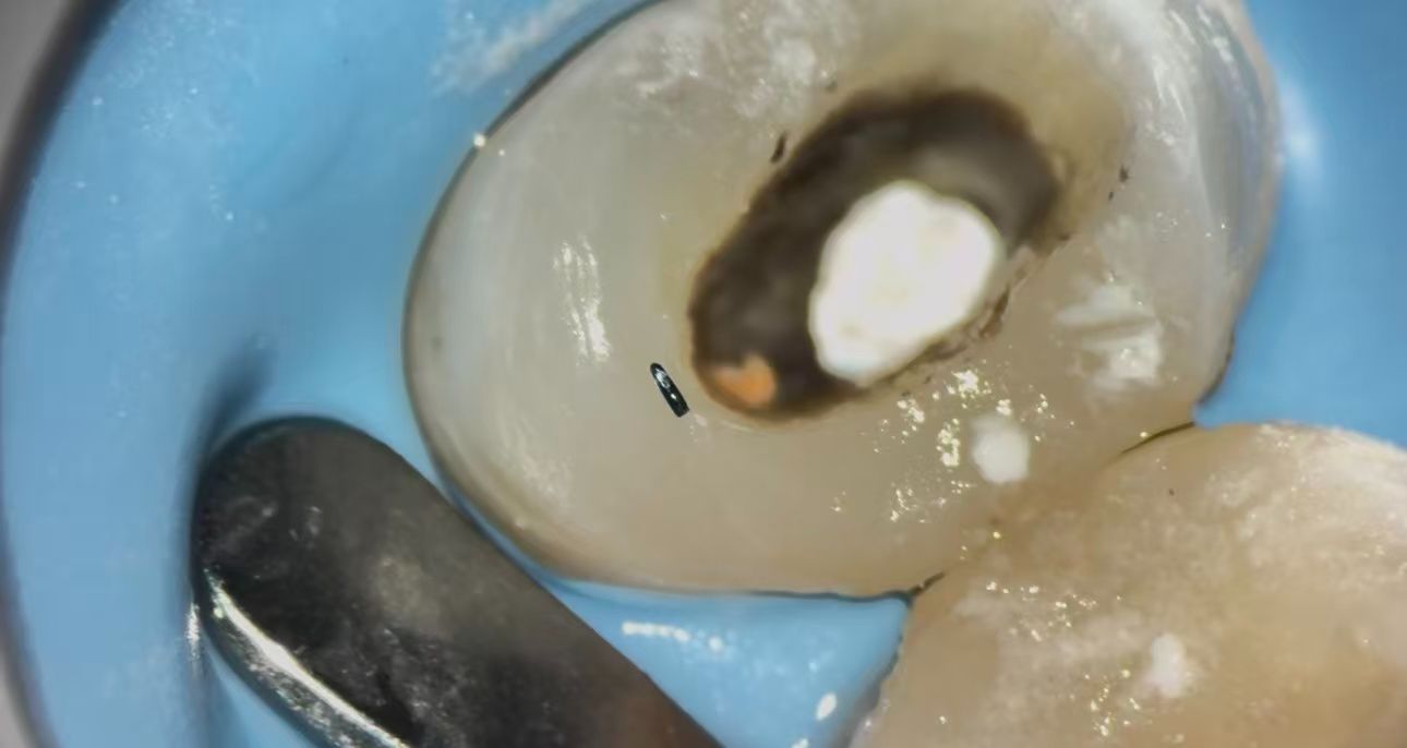
Access cavity refinement was performed under high magnification with a dental operating microscope to ensure optimal visibility and precision. The separated 2 mm instrument fragment was identified in the apical third of the buccal canal.
An ultrasonic E6 tip was applied with gentle, controlled vibration to conservatively trough dentin around the fragment and loosen it without excessive removal of tooth structure. Continuous microscopic monitoring allowed safe, atraumatic retrieval of the fragment without perforation or significant dentin loss. Magnification proved essential for preserving root integrity during this critical step.
Removal of Previous Obturation and Canal Preparation
Following fragment removal, the remaining obturation material was efficiently removed using Rogin retreatment rotary files, which effectively penetrated and cleared gutta-percha and sealer while respecting the original canal anatomy.
Subsequent shaping and refinement were accomplished with Rogin Super Flexi files. These heat-treated NiTi instruments offer:
- Exceptional flexibility
- High resistance to cyclic fatigue
- Excellent ability to maintain the natural canal curvature
- Suitability for retreatment cases and complex anatomies
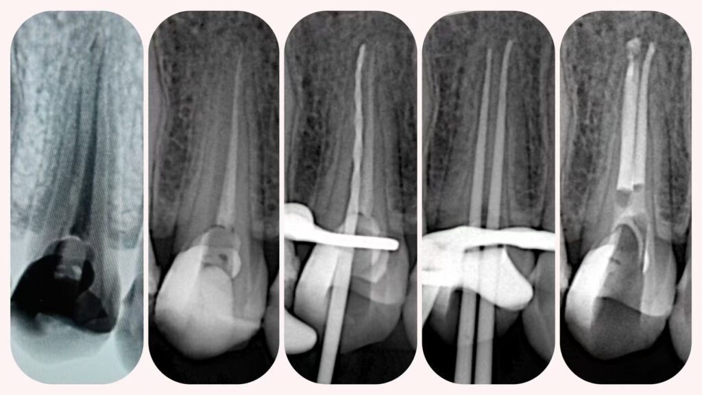
Working length was verified using an electronic apex locator.
Irrigation and Disinfection
Thorough disinfection is paramount in retreatment, particularly after instrument separation. The final irrigation protocol followed this sequence:
- EDTA (to remove the smear layer)
- Sodium hypochlorite (NaOCl)
- Chlorhexidine (for residual antimicrobial effect)
NaOCl irrigation was activated ultrasonically to enhance penetration into the apical third, lateral canals, and dentinal tubules, achieving more effective 3D cleaning of the root canal system.
Obturation
Once the canals were confirmed clean and dry, obturation was completed in the same visit using a 3D obturation technique with bioceramic sealer. This approach ensured complete filling of the canal space, including anatomical irregularities, and provided an excellent apical seal.
Coronal Seal and Follow-up
A reliable coronal seal was placed to prevent coronal leakage and reinfection. The patient was referred for definitive coronal restoration. Postoperative evaluation showed resolution of symptoms and no tenderness to percussion.
Discussion
Management of separated instruments requires a combination of advanced visualization (dental microscope), conservative ultrasonic techniques (e.g., E6 tip), and flexible rotary systems. Rogin retreatment files provided efficient removal of old fillings, while Super Flexi files allowed safe shaping without transportation or alteration of the natural canal pathway. Activated irrigation significantly enhanced cleaning efficacy in the apical region after fragment retrieval. Bioceramic sealer contributed to a dimensionally stable, biocompatible 3D obturation.
This case illustrates that even advanced retreatment scenarios involving apical-third separated instruments can be predictably resolved in a single session with proper armamentarium and meticulous technique.
Conclusion
Complex endodontic retreatments with separated instruments can achieve high success rates when supported by:
- Dental operating microscope for visualization
- Conservative ultrasonic retrieval techniques
- Effective, flexible rotary file systems (Rogin retreatment and Super Flexi)
- Activated irrigation protocols
- Modern 3D obturation with bioceramic sealers
Careful case selection, accurate diagnosis, appropriate tool selection, and precise execution remain the keys to success in challenging endodontic retreatment cases.