OpeningClinical Case by Dr. Nebras (Libya)
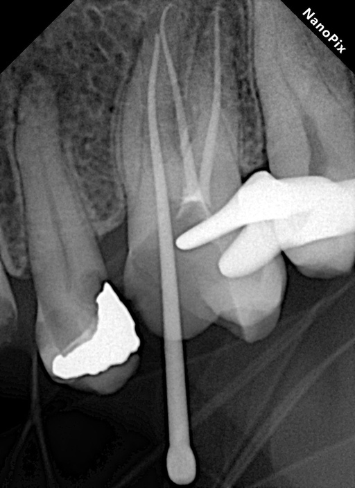
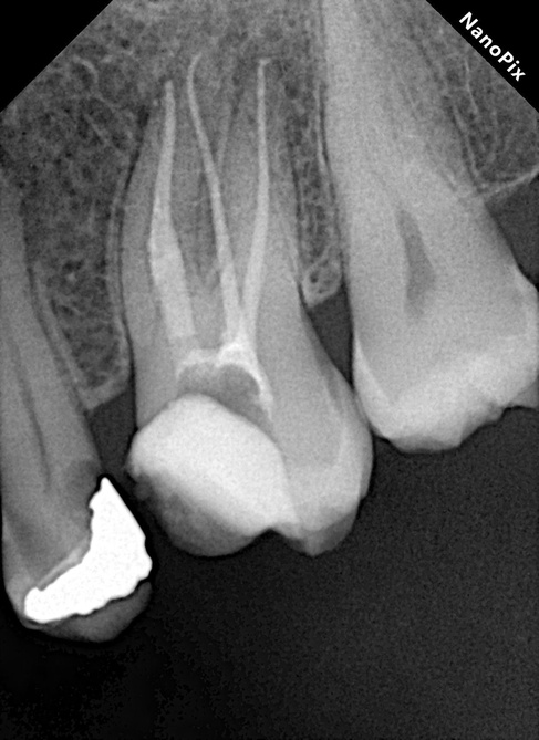
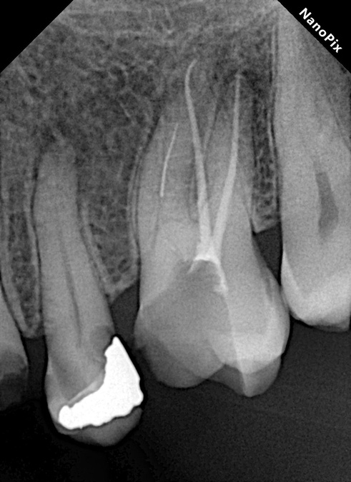
Complex root canal retreatments involving separated instruments remain one of the most demanding procedures in endodontics. When combined with limited mouth opening, the difficulty increases significantly. This clinical case demonstrates a systematic and minimally invasive approach to managing an asymptomatic apical abscess in tooth 26, complicated by a separated instrument in the mesiobuccal canal.
Case Details
- Tooth: 26 (Maxillary left first molar)
- Diagnosis: Asymptomatic apical abscess
- Primary Challenge: Retreatment with a 4.5 mm separated instrument located in the mesiobuccal canal, further complicated by limited mouth opening
- Case Difficulty: Advanced (Separated instrument retrieval & retreatment under restricted access)
Clinical Workflow
1. Access Cavity Refinement
The existing access cavity was carefully modified and orifice troughing was performed. Establishing a refined access and straight-line pathway is essential in patients with limited mouth opening, as it reduces instrument binding and minimizes fatigue during subsequent procedures.
2. Separated Instrument Retrieval
A modern wire-and-loop technique was employed under the dental operating microscope to retrieve the 4.5 mm separated fragment from the mesiobuccal canal. Notably, the retrieval was completed without creating a staging platform, helping preserve radicular dentin while achieving precise engagement of the fragment.
3. Glide Path Management
After successful retrieval, the canal was carefully scouted using a manual K-file #10 to confirm patency. This was followed by a rotary glide path file (#15/.03) to eliminate coronal interferences and create a smooth, secure pathway for further instrumentation.
4. Canal Preparation
Mechanical shaping was completed conservatively to a final size of #30/.04 using R-Shaper files. The instruments demonstrated outstanding efficiency, enabling controlled and predictable preparation while protecting the structural integrity of the root.
5. Canal Disinfection
A comprehensive irrigation protocol was applied using Sodium Hypochlorite, EDTA, and Chlorhexidine. Activation was performed prior to finalization to enhance the removal of residual debris and biofilm from the complex canal system.
6. Root Canal Obturation
Final obturation was carried out using a bioceramic sealer in combination with a warm (hot) obturation technique, ensuring a dense and homogeneous three-dimensional seal.
Clinical Tips
- Proper access refinement and orifice troughing are critical to minimize instrument binding and operator fatigue, especially in cases with limited mouth opening.
- High-magnification visualization under the microscope is indispensable for precise wire-and-loop engagement during separated instrument retrieval.
- Always begin with careful manual scouting using a #10 K-file to verify canal patency before introducing rotary pathfiles.
- Motorized glide path files effectively remove coronal interferences, allowing safer and more controlled progression into the canal.
- Maintaining a minimally invasive shaping protocol (final size #30/.04) helps safeguard long-term root structure integrity during complex retreatment cases.
- The R-Shaper files used in this case delivered excellent cutting efficiency and control, contributing significantly to the successful outcome.
Conclusion
This case highlights the value of a systematic, microscope-assisted approach when managing separated instruments in restricted clinical conditions. Through careful access refinement, the wire-and-loop retrieval technique, conservative glide path management, and efficient instrumentation with R-Shaper files, a predictable and dentin-preserving result was achieved.
Complex retreatments of this nature require both technical precision and the right instrumentation. When these elements are combined, even advanced cases involving limited mouth opening and intracanal obstacles can be managed successfully.