Root Canal Treatment of Maxillary Second Molar (Tooth 27) with Anxious Patient, Limited Mouth Opening, and Complex Canal Anatomy
Introduction
The endodontic management of complex root canal morphology is a demanding procedure in clinical dentistry. When combined with a dentally anxious patient and restricted mouth opening, the clinical challenge escalates significantly — demanding a file system that is not only highly flexible and safe, but also efficient and predictable.
This case report documents the successful nonsurgical root canal treatment of tooth 27 (upper left second molar) under precisely these demanding conditions, using the EZ Shaper Pro rotary file system by Rogin Dental. The case highlights how the EZ Shaper Pro system’s advanced heat-treated NiTi alloy, combined with a logical file sequence, enabled safe and efficient shaping of severely curved canals that would challenge conventional file systems.
Patient and Diagnosis
- Patient: 31 year old female with significant dental anxiety (managed with reassurance and careful communication throughout treatment)
- Chief Complaint: Spontaneous pain and sensitivity on palpation and percussion associated with the upper left second molar (tooth 27 / UL7)
- Radiographic Findings: CBCT and periapical radiographs revealed periapical pathology at tooth 27 with highly complex three-dimensional root canal anatomy
- Diagnosis: Pulp necrosis with symptomatic apical periodontitis (tooth 27)
Pre-operative Radiographic Imaging
Pre-operative CBCT imaging was essential to fully understand the three-dimensional anatomy of tooth 27 prior to treatment. The images clearly demonstrate the pronounced curvatures of the mesiobuccal (MB1) and distobuccal (DB) root canals — the key anatomical challenge in this case.
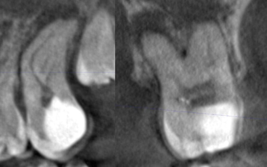
Fig. 1 — Pre-operative CBCT views of tooth 27, demonstrating pronounced multiplanar curvatures of the MB1 and DB canals and periapical pathology confirming the need for root canal treatment
Treatment Procedure
Access Cavity Preparation
A conservative access cavity was prepared under rubber dam isolation. Given the restricted mouth opening, access design was adapted to optimise straight-line access to all canal orifices. All three canal orifices (MB1, DB, and P) were identified and confirmed under magnification.
Biomechanical Preparation — EZ Shaper Pro System (Rogin Dental)
Canal shaping was performed exclusively using the EZ Shaper Pro rotary file system by Rogin Dental. This was the central instrument of this case, and its performance in the challenging MB1 and DB canals was the decisive factor in achieving a safe, efficient, and complication-free result.
The EZ Shaper Pro system by Rogin Dental enabled safe, efficient, and complication-free shaping of severely curved MB1 and DB canals — canals that represent the most challenging anatomy.
Why the EZ Shaper Pro Excelled in This Case
- The heat-treated NiTi alloy of the EZ Shaper Pro files allowed them to follow the natural curvature of the MB1 and DB canals without straightening, ledging, or transporting the canals.
- The advanced metallurgy of EZ Shaper Pro files provides enhanced resistance to cyclic fatigue failure, a key safety property when negotiating repeated, severe curvatures within a single canal preparation sequence.
- The EZ-1 file was used to perform initial coronal pre-flaring and establish a reproducible working taper in the demanding MB1 and DB canals. Its performance in these canals was notably smooth and controlled — characteristics that are particularly valued in cases where repeated file cycling over tight curvatures could otherwise cause fatigue failure.
- Following EZ-1 pre-flaring, the remaining EZ Shaper Pro files were used in a sequential crown-down sequence, progressively preparing the middle and apical thirds of each canal. The logical, graduated file sequence allowed precise control over the shaping process.
- The EZ Shaper Pro system accommodated the different anatomical requirements of this molar — the MB1 and DB canals (smaller, more curved) were shaped to 25/.04, while the larger, straighter palatal canal was efficiently shaped to 35/.03, demonstrating the system’s wide clinical range.
At no point during the shaping procedure was there any sign of procedural error, instrument binding, or deviation from the pre-operative canal path. The EZ Shaper Pro files tracked the canal curvature faithfully throughout, confirming the reliability and safety of the system in this challenging case.
Intraoral Photographs — Following Shaping (Pre-obturation)
The photograph below, taken under rubber dam isolation and magnification following completion of canal shaping, shows the three prepared canal orifices of tooth 27.
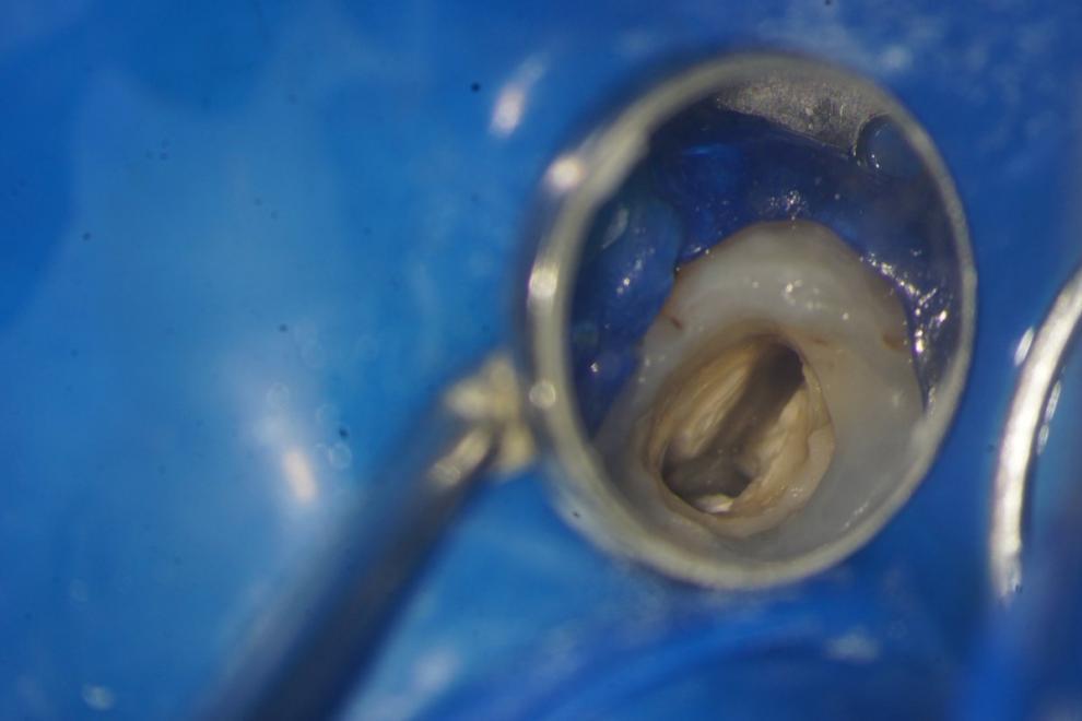
Fig. 2 — Intraoral photograph following EZ Shaper Pro canal preparation (pre-obturation). Three clearly defined, well-shaped orifices are visible (MB1, DB, P), demonstrating the conservative and controlled shaping achieved by the EZ Shaper Pro system.
Irrigation and Disinfection
Irrigation Protocol
- 5.25% sodium hypochlorite (NaOCl) — primary irrigant used throughout shaping to dissolve organic tissue and provide continuous disinfection
- 17% EDTA — applied following canal shaping to remove the smear layer and open dentinal tubules for enhanced sealer penetration
- Final flush with distilled water — to clear residual irrigants prior to obturation
Rogin Dental Flexible Irrigation Needle
All irrigants were delivered using Rogin Dental’s flexible polymer irrigation needle — a closed-tip, side-vented, angled needle engineered to deliver irrigants safely and precisely to the apical third of curved root canals. The needle’s flexible polymer body allowed it to follow the curvature of the MB1 and DB canals without risk of perforation or ledging, while the side-vent design ensured controlled, low-pressure irrigant delivery with no risk of apical extrusion.
This needle is a natural companion to the EZ Shaper Pro system: both are engineered by Rogin Dental to work with curved canal anatomy rather than against it, combining to deliver thorough chemo-mechanical debridement even in the most challenging root canal systems.
Obturation
Following canal drying with appropriately sized paper points, obturation was performed using the hydraulic condensation technique in combination with WellRoot ST bioceramic sealer. The hydraulic condensation approach was selected specifically for its ability to drive the highly flowable bioceramic sealer three-dimensionally through the complex canal system, including any lateral canals, fins, or anastomoses present in this anatomically demanding molar.
The combination of conservative EZ Shaper Pro preparation (preserving dentinal walls while achieving adequate shape) and hydraulic bioceramic obturation represents an optimal approach for cases of this complexity — ensuring structural integrity of the root while achieving a predictable, hermetic seal.
Intraoral Photographs — Post-obturation
The photograph below was taken immediately following completion of hydraulic condensation obturation, prior to coronal restoration. The obturated canal orifices are clearly visible, with gutta percha points confirmed within all three canal systems.
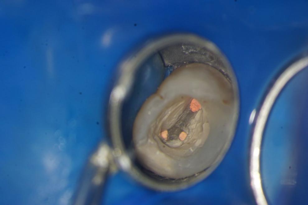
Fig. 3 — Intraoral photograph immediately following hydraulic condensation obturation with WellRoot ST bioceramic sealer. All three EZ Shaper Pro-prepared canals (MB1, DB, P) are fully obturated.
Post-operative Assessment
Post-operative Radiographic Evaluation
Post-operative CBCT and periapical radiographs confirmed homogeneous, well-condensed obturation material within all three root canal systems (MB1, DB, and P), with the fill extending to the established working lengths. The radiographic appearance is consistent with a thorough three-dimensional seal achieved by hydraulic condensation with WellRoot ST bioceramic sealer. The canal paths visible on post-operative imaging confirm faithful preservation of the original canal anatomy — direct evidence of the shape-preserving properties of the EZ Shaper Pro system.
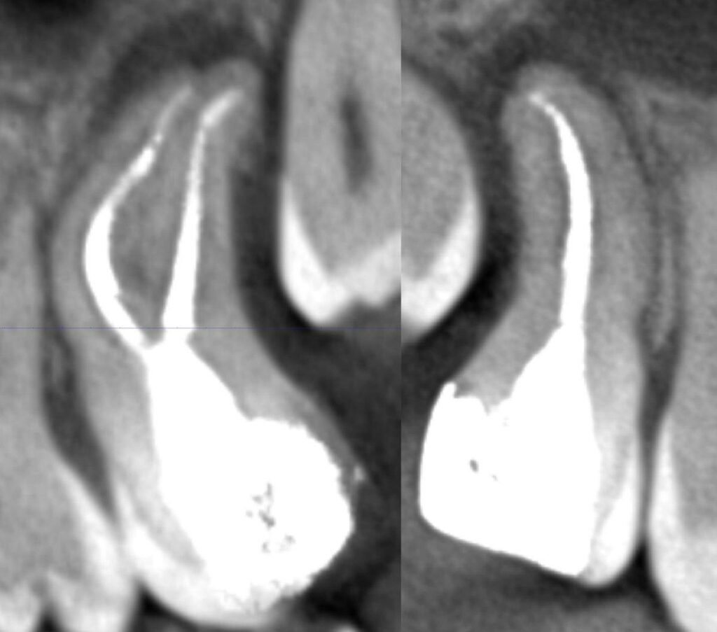
Fig. 4 — Post-operative CBCT views of tooth 27 following obturation. All three canals are densely filled to working length, confirming the quality of the EZ Shaper Pro preparation and hydraulic condensation obturation.
Follow-up radiographic monitoring is planned at 3, 6, and 12 months to assess periapical healing.
Conclusion
This case demonstrates the exceptional clinical performance of the EZ Shaper Pro rotary file system (Rogin Dental) in one of endodontics’ most challenging scenarios: a severely curved, multi-rooted maxillary molar in an anxious patient with limited mouth opening. The EZ Shaper Pro system — anchored by the versatile EZ-1 file for initial coronal flaring and progressing through a logical, graduated sequence — delivered safe, efficient, and complication-free preparation of the MB1 and DB canals despite their pronounced multiplanar curvatures.
EZ Shaper Pro‘s heat-treated NiTi alloy, superior flexibility, and cyclic fatigue resistance make it the file system of choice for curved, complex canal anatomy — as proven in this demanding clinical case.Combined with Rogin Dental’s flexible irrigation needle for thorough chemo-mechanical debridement and the EZ Shaper Pro system enabled a fully predictable, high-quality outcome. This case stands as a compelling example of what becomes achievable — even in the most demanding clinical conditions — when the right instruments are used.
