Root canal treatment on a mandibular first molar can present significant challenges, especially when intricate canal anatomy and severe narrowing are involved. In this detailed endodontic case report, Dr. Nebras shares a successful approach to treating Tooth #46 diagnosed with a symptomatic apical abscess. This case highlights the importance of advanced techniques and specialized instruments in managing complex root canal anatomy.
Keywords: endodontic treatment, mandibular molar root canal, Vertucci classification, symptomatic apical abscess, microscopic endodontics
Case Overview
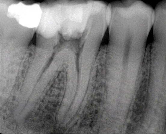
Tooth: Mandibular 1st Molar (#46)
Diagnosis: Symptomatic apical abscess
This case involved notable anatomical complexities that demanded precision and careful instrumentation:
- Vertucci Class VI configuration in the distal canal
- Vertucci Class II configuration in the mesial canals
- Severely narrowed mesial canals, increasing the risk of file separation
Such variations in root canal anatomy are common in mandibular molars and require a meticulous, magnification-assisted approach to ensure thorough cleaning, shaping, and disinfection while minimizing procedural errors.
Clinical Challenges in Complex Endodontics
Vertucci Class VI (distal canal) and Vertucci Class II (mesial canals) represent challenging configurations that can complicate negotiation, glide path creation, and biomechanical preparation. Combined with severely calcified or narrowed mesial canals, these factors significantly elevate the risk of iatrogenic errors such as ledge formation or instrument separation.
Successful management relies on:
- Enhanced visualization through the dental operating microscope
- Ultrasonic assistance for precise access
- Highly flexible and efficient rotary files
- Controlled tactile feedback during instrumentation
Detailed Clinical Workflow
The treatment followed a systematic, magnification-enhanced protocol:
1. Access Cavity Preparation
- Microscopic management was employed throughout the procedure for superior visualization.
- Access was modified using ultrasonic troughing to locate and deroof all canals effectively.
2. Canal Negotiation and Patency
- Initial negotiation and patency were achieved with size 10 manual K-files.
- This step was critical in the severely narrowed mesial canals to establish a safe pathway.
3. Glide Path Creation
- A Rogin Path File 15/.03 was used to create a reliable glide path, ensuring smooth transition to rotary instrumentation.
4. Biomechanical Preparation
- Canals were prepared under magnification using Aurora Rogin Super Flexi files.
- Sequence: 20/.04 → 25/.04 → 30/.04
- Copious irrigation was performed between each step to maintain canal patency and remove debris.
5. Final Irrigation Protocol
- A comprehensive final rinsing protocol was completed with ultrasonic activation to enhance disinfection in complex anatomies.
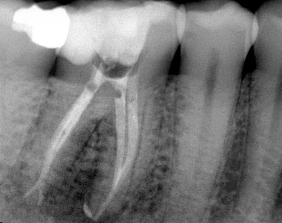
6. Obturation
- The canals were obturated using hot obturation techniques for optimal three-dimensional sealing.
Why Aurora Rogin Super Flexi Files Excelled in This Case
Dr. Nebras specifically utilized Aurora Rogin Super Flexi files for instrumentation. These files demonstrated:
- Exceptional cutting efficiency
- Outstanding flexibility — essential for navigating Vertucci Class VI and Class II configurations
- Superior performance in severely narrowed canals
The combination of flexibility and controlled cutting allowed safe and effective shaping without compromising the original canal anatomy.
Clinical Tips for Managing Complex Root Canal Cases
- Tactile control activation with small rotary files is crucial in severely narrowed canals to prevent procedural errors.
- In cases with complex Vertucci anatomy, small, highly flexible rotary files are non-negotiable.
- Microscopic visualization combined with ultrasonic troughing significantly improves canal localization and reduces missed anatomy.
- Always maintain copious irrigation and use activation techniques for optimal cleaning in multi-canal teeth with anatomical variations.
Conclusion
This endodontic case report demonstrates that even advanced challenges — such as symptomatic apical abscess in a mandibular first molar with complex Vertucci classifications and narrowed canals — can be managed predictably with proper technique, magnification, and the right instruments.
The Aurora Rogin Super Flexi files proved invaluable in achieving safe negotiation and preparation. For endodontists and general practitioners handling complex root canal treatments, investing in advanced visualization and flexible rotary systems can dramatically improve outcomes.
Dr. Nebras specializes in challenging endodontic cases. This case highlights the power of modern technology and technique in preserving natural teeth.