Managing calcified canals combined with a periapical lesion represents one of the more demanding challenges in endodontics. This advanced clinical case from Iraq, treated by Dr. Abdulrahman Tariq, demonstrates a systematic approach to negotiating complete canal calcification in tooth #25 while achieving healing of the associated periapical pathology.
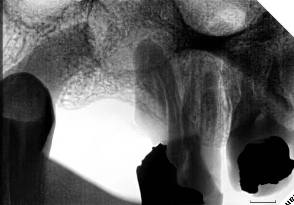
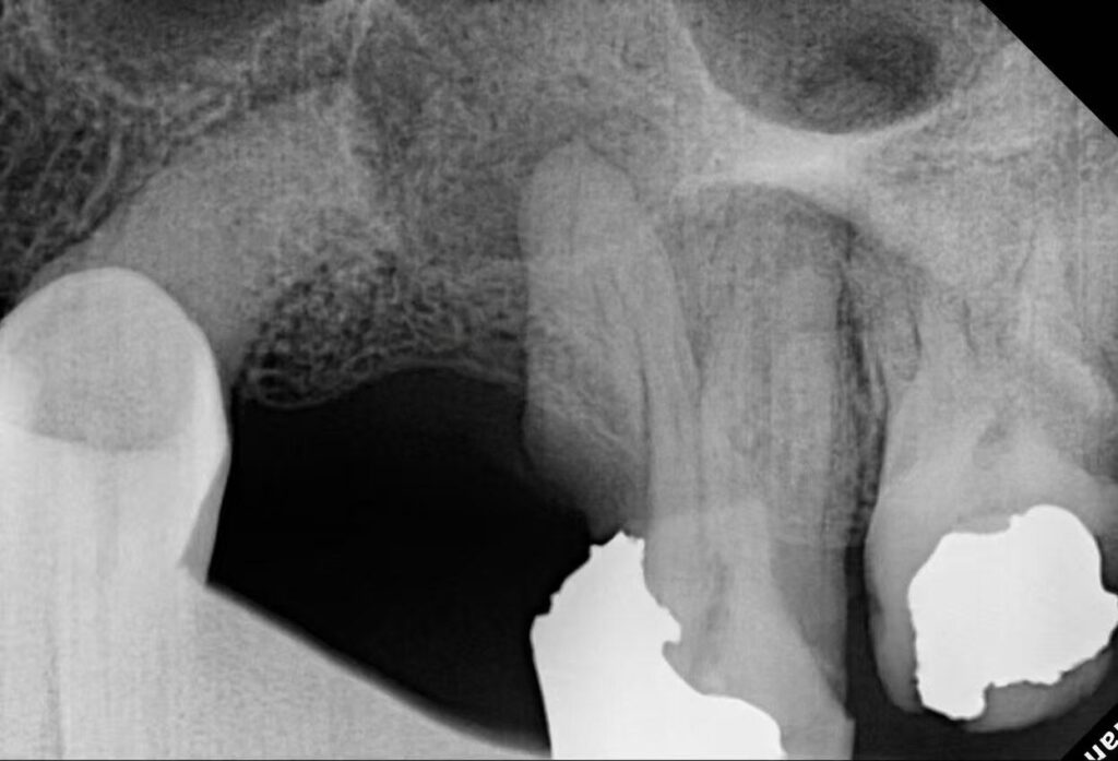
Case Presentation
A patient presented with severe pain and swelling in the upper left region. Clinical and radiographic examination revealed that tooth #25 exhibited a clear periapical lesion together with complete calcification of the root canal system. Root canal therapy was indicated to resolve both the pulpal pathology and the apical disease.
Case Difficulty: Advanced
Tooth: #25 (upper left second premolar)
Diagnosis: Pulpal pathology + Periapical lesion with complete canal calcification
Primary Challenges: Complete canal calcification and Vertucci Type II anatomy
Clinical Challenges
Complete calcification significantly increases the difficulty of locating and negotiating the canal system. In addition, the tooth presented Vertucci Type II anatomy (two canals joining into one), which requires careful sequential instrumentation to maintain patency and avoid ledging or perforation. The presence of an established periapical lesion further underscored the need for thorough cleaning, shaping, and hermetic obturation.
Clinical Workflow
1. Access Opening
Access was prepared through the existing crown, allowing direct entry into the pulp chamber while preserving as much coronal structure as possible.
2. Establishing Patency
The key to successfully opening and patenting the completely calcified canals was the sequential use of EZx followed by EZ1, performed under copious irrigation. Continuous irrigation during negotiation prevented debris accumulation and blockage, enabling progressive advancement into the calcified space until full working length was reached.
3. Canal Instrumentation
Once patency was confirmed, the canals were shaped using the EZ sequence: EZ2 → EZ3 → EZ4. This progressive enlargement respected the Vertucci Type II configuration, allowing controlled preparation of both canals that ultimately joined apically.
4. Irrigation Protocol
A comprehensive irrigation protocol with activation was carried out to enhance chemical disinfection throughout the complex canal anatomy. Activation improves irrigant penetration into residual irregularities and the apical third, which is particularly important in previously calcified systems.
5. Obturation
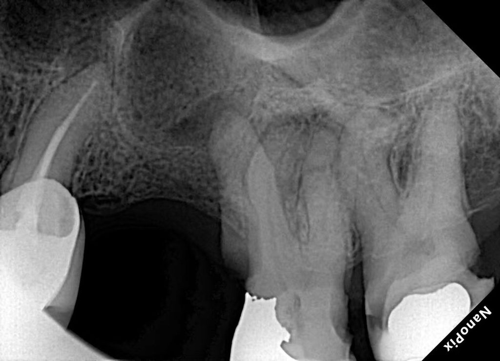
Obturation was performed using a modified warm vertical compaction technique. This method provided dense, three-dimensional filling of the prepared canal space, including the junction area characteristic of Vertucci Type II anatomy.
6. Follow-up
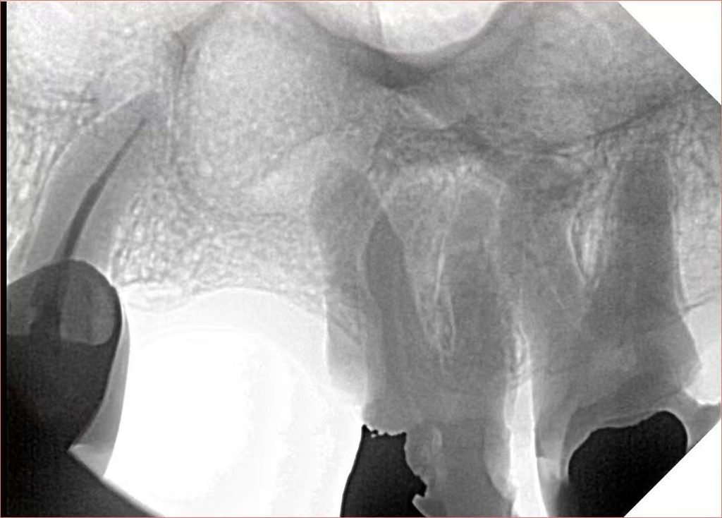
A 6-month postoperative radiograph demonstrated clear healing of the periapical lesion, confirming the biological success of the treatment despite the initial complete calcification.
Clinical Tips from Dr. Abdulrahman Tariq
- EZx and EZ1 are decisive instruments for negotiating completely calcified canals; their controlled cutting ability allows safe advancement where conventional files may fail.
- Maintain copious irrigation throughout the patency phase to prevent debris blockage and maintain a clear pathway.
- In Vertucci Type II anatomy, progress gently and sequentially through the EZ2–EZ3–EZ4 sequence to avoid separating or transporting the canals.
- Radiographic evidence of periapical lesion healing at 6 months remains a reliable indicator of treatment success, even in cases that began with severe calcification.
Conclusion
This advanced case illustrates that complete canal calcification, when approached with a systematic protocol centered on EZx and EZ1 for initial negotiation, followed by controlled shaping with the EZ sequence, thorough irrigation, and precise obturation, can lead to predictable clinical and radiographic success. The 6-month healing of the periapical lesion confirms that even highly challenging anatomy can be managed effectively when the correct instruments and technique are applied.
Careful case assessment, patience during canal negotiation, and respect for complex anatomical variations such as Vertucci Type II remain fundamental to achieving long-term endodontic success in calcified teeth.
